Citation:
| 1.09 MB |
Date Published:
Sep 15Abstract:
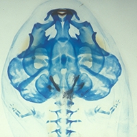
The contribution of cranial neural crest cells to the development and patterning of cranial muscles in amphibians was investigated in the phylogenetically basal and morphologically generalized frog, Bombina orientalis. Experimental methods included fluorescent marking of premigratory cranial neural crest and extirpation of individual migratory streams. Neural crest cells contributed to the connective tissue component, but not the myofibers, of many larval muscles within the first two branchial arches (mandibular and hyoid), and complex changes in muscle patterning followed neural crest extirpation. Connective tissue components of individual muscles of either arch originate from the particular crest migratory stream that is associated with that arch, and this relationship is maintained regardless of the segmental identity-or embryonic derivation-of associated skeletal components. These developmental relations define a pattern of segmentation in the head of larval anurans that is similar to that previously described in the domestic chicken, the only vertebrate that has been thoroughly investigated in this respect. The fundamental role of the neural crest in patterning skeleton and musculature may represent a primitive feature of cranial development in vertebrates. Moreover, the corresponding developmental processes and cell fates appear to be conserved even when major evolutionary innovations-such as the novel cartilages and muscles of anuran larvae-result in major differences in cranial form. (C) 2001 Academic Press.
Notes:
476CQTimes Cited:48Cited References Count:47