Journal Articles and Book Chapters: 2009 - 2000

2009
Wu, Y.K., et al., 2009. A new species of the genus Paramesotriton (Caudata: Salamandridae) from Guangxi Zhuang Autonomous Region, southern China. Zootaxa , pp. 59-68.Abstract

A new species of Asian warty newt, Paramesotriton ermizhaoi, is described from Guangxi Zhuang Autonomous Region in southern China. This species is easily distinguished from other congeners by external morphology and osteology. Phylogenetic analyses based on mitochondrial data place P. ermizhaoi as a possible sister taxon to P. hongkongensis and P. chinensis. The number of trunk vertebrae varies from 12 to 13 in the new species. The relationship between P. ermizhaoi and a potentially new Pachytriton species (Pachytriton C), and the Guangxi population of P. chinensis is briefly discussed.

PDF
2008
Meegaskumbura, M., et al., 2008. Pickled frogs help biodiversity assessment. In S. Stuart, et al., ed. Threatened Amphibians of the World. Barcelona, Gland, Arlington. Barcelona, Gland, Arlington: Lynx Edicions, IUCN, and Conservation International, pp. 45. PDF
Blackburn, D.C., Hanken, J. & Jenkins, F.A., 2008. Concealed weapons: erectile claws in African frogs. Biology Letters , 4 , pp. 355-357.Abstract

Vertebrate claws are used in a variety of important behaviours and are typically composed of a keratinous sheath overlying the terminal phalanx of a digit. Keratinous claws, however, are rare in living amphibians; their microstructure and other features indicate that they probably originated independently from those in amniotes. Here we show that certain African frogs have a different type of claw, used in defence, that is unique in design among living vertebrates and lacks a keratinous covering. These frogs have sectorial terminal phalanges on their hind feet that become functional by cutting through the skin. In the resting state, the phalanx is subdermal and attached to a distal bony nodule, a neomorphic skeletal element, via collagen-rich connective tissue. When erected, the claw breaks free from the nodule and pierces the ventral skin. The nodule, suspended by a sheath attached to the terminal phalanx and supported by collagenous connections to the dermis, remains fixed in place. While superficially resembling the shape of claws in other tetrapods, these are the only vertebrate claws known to pierce their way to functionality.

PDF
Zimmermann, J., et al., 2008. DNA damage in preserved specimens and tissue samples: a molecular assessment. Frontiers in Zoology , 5.Abstract

The extraction of genetic information from preserved tissue samples or museum specimens is a fundamental component of many fields of research, including the Barcode of Life initiative, forensic investigations, biological studies using scat sample analysis, and cancer research utilizing formaldehyde-fixed, paraffin-embedded tissue. Efforts to obtain genetic information from these sources are often hampered by an inability to amplify the desired DNA as a consequence of DNA damage. Previous studies have described techniques for improved DNA extraction from such samples or focused on the effect of damaging agents - such as light, oxygen or formaldehyde - on free nucleotides. We present ongoing work to characterize lesions in DNA samples extracted from preserved specimens. The extracted DNA is digested to single nucleosides with a combination of DNase I, Snake Venom Phosphodiesterase, and Antarctic Phosphatase and then analyzed by HPLC-ESI-TOF-MS. We present data for moth specimens that were preserved dried and pinned with no additional preservative and for frog tissue samples that were preserved in either ethanol, or formaldehyde, or fixed in formaldehyde and then preserved in ethanol. These preservation methods represent the most common methods of preserving animal specimens in museum collections. We observe changes in the nucleoside content of these samples over time, especially a loss of deoxyguanosine. We characterize the fragmentation state of the DNA and aim to identify abundant nucleoside lesions. Finally, simple models are introduced to describe the DNA fragmentation based on nicks and double-strand breaks.

PDF
Kerney, R. & Hanken, J., 2008. Gene expression reveals unique skeletal patterning in the limb of the direct-developing frog, Eleutherodactylus coqui. Evolution & Development , 10 , pp. 439-448.Abstract

The growing field of skeletal developmental biology provides new molecular markers for the cellular precursors of cartilage and bone. These markers enable resolution of early features of skeletal development that are otherwise undetectable through conventional staining techniques. This study investigates mRNA distributions of skeletal regulators runx2 and sox9 along with the cartilage-dominant collagen 2(alpha)1 (col2a1) in embryonic limbs of the direct-developing anuran, Eleutherodactylus coqui. To date, distributions of these genes in the limb have only been examined in studies of the two primary amniote models, mouse and chicken. In E coqui, expression of transcription factors runx2 and sox9 precedes that of col2a1 by 0.5-1 developmental stage (approximately 12-24 h). Limb buds of E. coqui contain unique distal populations of both runx2- and sox9-expressing cells, which appear before formation of the primary limb axis and do not express col2a1. The subsequent distribution of col2a1 reveals a primary limb axis similar to that described for Xenopus laevis. Precocious expression of both runx2 and sox9 in the distal limb bud represents a departure from the conserved pattern of proximodistal formation of the limb skeleton that is central to prevailing models of vertebrate limb morphogenesis. Additionally, runx2 is expressed in the early joint capsule perichondria of the autopod and in the perichondria of long bones well before periosteum formation. The respective distributions of sox9 and col2a1 do not reveal the joint perichondria but instead are expressed in the fibrocartilage that fills each presumptive joint capsule. These distinct patterns of runx2- and sox9-expressing cells reveal precursors of chondrocyte and osteoblast lineages well before the appearance of mature cartilage and bone.

PDF
Gross, J.B. & Hanken, J., 2008. Review of fate-mapping studies of osteogenic cranial neural crest in vertebrates. Developmental Biology , 317 , pp. 389-400.Abstract

Recent years have witnessed renewed interest in defining the embryonic cell populations that directly contribute to the bony skull. This question lies at the intersection of several important developmental, clinical and evolutionary interests. Until recently, our collective understanding of the embryonic origin of the vertebrate osteocranium has been based on a small number of reports published solely using avian models. As data gradually accumulates from other, distantly related species (e.g., mouse and frog), we can begin to evaluate long-standing assumptions regarding the behavior of osteogenic (bone-forming) neural crest cells within a wider phylogenetic and comparative context. In this review, we summarize data collected to date in three major vertebrate taxa: amphibians, birds and mammals. We highlight three largely unexplored topics within the field of osteogenic neural crest development: 1) disagreements in bone tissue origin within and across current model systems; 2) whether the pattern of neural crest cell contribution to skull bone is evolutionarily conservative or labile; and 3) how our understanding of development and morphology will benefit from fate maps using currently unexamined animal models. (C) 2008 Elsevier Inc. All rights reserved.

PDF
Gross, J.B. & Hanken, J., 2008. Segmentation of the vertebrate skull: neural-crest derivation of adult cartilages in the clawed frog, Xenopus laevis. Integrative and Comparative Biology , 48 , pp. 681-696.Abstract

We utilize a novel, transgenic cell-labeling system to assess the embryonic derivation of cartilages in the post-metamorphic skull of anuran amphibians. Many of these cartilages form de novo at metamorphosis and have no obvious precursors within the larval skeleton. Most adult cartilages are derived from mandibular- or hyoid-stream neural crest, either individually or in combination; branchial-stream neural crest makes a modest contribution. Each stream also contributes to at least one cartilage in the middle ear or external ear. Four cartilages are composite elements; each is derived from at least two distinct cell populations. Many boundaries between adjacent neural-crest territories are cryptic insofar as they do not coincide with anatomical boundaries. The system of adult cranial segmentation revealed by these fate-mapping results differs in important respects from both the segmentation of the ontogenetically earlier larval skull and the cranial segmentation in amniotes. Most striking is the rostral inversion of neural-crest-derived cartilages in Xenopus, such that mandibular stream-derived elements are deployed caudal to those derived from the hyoid stream, which predominate anteriorly. This novel pattern of rostral segmentation may be a consequence of the complex, biphasic life history that is characteristic of most species of living amphibians, and especially anurans, in which cranial architecture is significantly reconfigured at metamorphosis. Neural-crest derivation of the vertebrate skull is not invariant; instead, embryonic derivation of individual components of the cranial skeleton may vary widely among species.

PDF
2007
Parra, G., et al., 2007. Systematics and Conservation. In C. Gascon, et al., ed. Amphibian Conservation Action Plan. Gland and Cambridge. Gland and Cambridge: IUCN/SSC Amphibian Specialist Group, pp. 45-48. PDF
Kerney, R., et al., 2007. 
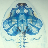
Cranial ontogeny in Philautus silus (Anura : Ranidae : Rhacoohorinae) reveals few similarities with other direct-developing anurans. Journal of Morphology , 268 , pp. 715-725.Abstract

Direct development has evolved in rhacophorine frogs independently from other anuran lineages, thereby offering an opportunity to assess features associated with this derived life history. Using a developmental series of the direct-developing Philautus silus (Ranidae: Rhaeophorinae) from Sri Lanka, we examine features of cranial morphology that are part of a suite of adaptations that facilitate feeding in free-living tadpoles, but have been changed or lost in other direct-developing lineages. Larval-specific upper jaw cartilages, which are absent from many non-rhacophorine direct-developing species (such as Eleutherodactylus coqui), develop in embryos of P. silus. Similarly, lower jaw cartilages initially assume a larval morphology, which is subsequently remodeled into the adult jaw configuration before hatching. However, the cartilaginous jaw suspension and hyobranchial skeleton never assume a typical larval morphology. The palatoquadrate, which suspends the lower jaw, lacks the posterior connections to the braincase found in many metamorphosing species. Unlike in metamorphosing species, bone formation in P. silus begins before hatching. However, the sequence of bone formation resembles that of metamorphosing anurans more than that of other direct developers. In particular, P. silus does not exhibit precocious ossification of the lower jaw, which is characteristic of some frogs and caecilians that lack a free-living tadpole. These data reveal some similarities between Philautus and other direct-developing anurans. However, the departure of Philautus embryos from the generalized tadpole skeletal morphology is less pronounced than that observed in other direct-developing taxa.

PDF
Kerney, R., Gross, J.B. & Hanken, J., 2007. Runx2 is essential for larval hyobranchial cartilage formation in Xenopus laevis. Developmental Dynamics , 236 , pp. 1650-1662.Abstract

The vertebrate transcription factor protein Runx2 is regarded as a "master regulator" of bone formation due to the dramatic loss of the osseous skeleton in the mouse homozygous knockout. However, Runx2 mRNA also is expressed in the pre-hypertrophic cartilaginous skeleton of the mouse and chicken, where its developmental function is largely unknown. Several tiers of Runx2 regulation exist in the mouse, any of which may account for its seeming biological inactivity during early stages of skeletogenesis. Unlike mouse and chicken, zebrafish require Runx2 function in early cartilage differentiation. The present study reveals that the earlier functional role of Runx2 in cartilage differentiation is shared between zebrafish and Xenopus. A combination of morpholino oligonucleotide injections and neural crest transplants indicate that Runx2 is involved in differentiation of the cartilaginous hyobranchial skeleton in the frog, Xenopus laevis. Additionally, in situ hybridizations show runx2 mRNA expression in mesenchymal precursors of the cartilaginous skull, which reveals the earliest pre-patterning of these cartilages described to date. The early distribution of runx2 resolves the homology of the larval suprarostral plate, which is one of the oldest controversies of anuran skull development. Together these data reveal a shift in Runx2 protein function during vertebrate evolution towards its exclusive roles in cartilage hypertrophy and bone differentiation within the amniote lineage.

PDF
Bininda-Emonds, O.R.P., et al., 2007. Forelimb-hindlimb developmental timing changes across tetrapod phylogeny. Bmc Evolutionary Biology , 7 , pp. 182.Abstract

Background: Tetrapods exhibit great diversity in limb structures among species and also between forelimbs and hindlimbs within species, diversity which frequently correlates with locomotor modes and life history. We aim to examine the potential relation of changes in developmental timing (heterochrony) to the origin of limb morphological diversity in an explicit comparative and quantitative framework. In particular, we studied the relative time sequence of development of the forelimbs versus the hindlimbs in 138 embryos of 14 tetrapod species spanning a diverse taxonomic, ecomorphological and life-history breadth. Whole- mounts and histological sections were used to code the appearance of 10 developmental events comprising landmarks of development from the early bud stage to late chondrogenesis in the forelimb and the corresponding serial homologues in the hindlimb.Results: An overall pattern of change across tetrapods can be discerned and appears to be relatively clade- specific. In the primitive condition, as seen in Chondrichthyes and Osteichthyes, the forelimb/ pectoral fin develops earlier than the hindlimb/ pelvic fin. This pattern is either retained or re- evolved in eulipotyphlan insectivores (= shrews, moles, hedgehogs, and solenodons) and taken to its extreme in marsupials. Although exceptions are known, the two anurans we examined reversed the pattern and displayed a significant advance in hindlimb development. All other species examined, including a bat with its greatly enlarged forelimbs modified as wings in the adult, showed near synchrony in the development of the fore and hindlimbs.Conclusion: Major heterochronic changes in early limb development and chondrogenesis were absent within major clades except Lissamphibia, and their presence across vertebrate phylogeny are not easily correlated with adaptive phenomena related to morphological differences in the adult fore- and hindlimbs. The apparently conservative nature of this trait means that changes in chondrogenetic patterns may serve as useful phylogenetic characters at higher taxonomic levels in tetrapods. Our results highlight the more important role generally played by allometric heterochrony in this instance to shape adult morphology.

PDF
Wake, D.B., Savage, J.M. & Hanken, J., 2007. Montane salamanders from the Costa Rica-Panama border region, with descriptions of two new species of Bolitoglossa. Copeia , pp. 556-565.Abstract

Two new species of lungless salamanders (Plethodontidae) are described from montane habitats of eastern Costa Rica and adjacent western Panama. Bolitoglossa gomezi and B. bramei are distinguished from each other and from other salamander species in this remote area in adult body size, external proportions, foot webbing, tooth counts, and/or external coloration. Both new species are assigned to the B. subpalmata species group, subgenus Eladinea. A newly identified specimen of Bolitoglossa anthracina-only the fourth known specimen of this rare species in collections-is reported from the same region. Salamander species diversity along the border between Costa Rica and Panama is exceptionally large, at present comprising 22 named and two unnamed forms.

PDF
2006
Gross, J.B., et al., 2006. Use of a ROSA26 : GFP transgenic line for long-term Xenopus fate-mapping studies. Journal of Anatomy , 209 , pp. 401-413.Abstract

Widespread and persistent marker expression is a prerequisite for many transgenic applications, including chimeric transplantation studies. Although existing transgenic tools for the clawed frog, Xenopus laevis, offer a number of promoters that drive widespread expression during embryonic stages, obtaining transgene expression through metamorphosis and into differentiated adult tissues has been difficult to achieve with this species. Here we report the application of the murine ROSA26 promoter in Xenopus. GFP is expressed in every transgenic tissue and cell type examined at post-metamorphic stages. Furthermore, transgenic ROSA26:GFP frogs develop normally, with no apparent differences in growth or morphology relative to wild-type frogs. ROSA26 transgenes may be used as a reliable marker for embryonic fate-mapping of adult structures in Xenopus laevis. Utility of this transgenic line is illustrated by its use in a chimeric grafting study that demonstrates the derivation of the adult bony jaw from embryonic cranial neural crest.

PDF
2005
Gross, J.B. & Hanken, J., 2005. Cranial neural crest contributes to the bony skull vault in adult Xenopus laevis: Insights from cell labeling studies. Journal of Experimental Zoology Part B-Molecular and Developmental Evolution , 304B , pp. 169-176.Abstract

As a step toward resolving the developmental origin of the ossified skull in adult anurans, we performed a series of cell labeling and grafting studies of the cranial neural crest (CNC) in the clawed frog, Xenopus laevis. We employ an indelible, fixative-stable fluorescent dextran as a cell marker to follow migration of the three embryonic streams of cranial neural crest and to directly assess their contributions to the bony skull vault, which forms weeks after hatching. The three streams maintain distinct boundaries in the developing embryo. Their cells proliferate widely through subsequent larval (tadpole) development, albeit in regionally distinct portions of the head. At metamorphosis, each stream contributes to the large frontoparietal bone, which is the primary constituent of the skull vault in adult anurans. The streams give rise to regionally distinct portions of the bone, thereby preserving their earlier relative position anteroposteriorly within the embryonic neural ridge. These data, when combined with comparable experimental observations from other model species, provide insights into the ancestral pattern of cranial development in tetrapod vertebrates as well as the origin of differences reported between birds and mammals. (c) 2005 Wiley-Liss, Inc.

PDF
Hanken, J. & Gross, J.B., 2005. Evolution of cranial development and the role of neural crest: insights from amphibians. Journal of Anatomy , 207 , pp. 437-446.Abstract

Contemporary studies of vertebrate cranial development document the essential role played by the embryonic neural crest as both a source of adult tissues and a locus of cranial form and patterning. Yet corresponding and basic features of cranial evolution, such as the extent of conservation vs. variation among species in the contribution of the neural crest to specific structures, remain to be adequately resolved. Investigation of these features requires comparable data from species that are both phylogenetically appropriate and taxonomically diverse. One key group are amphibians, which are uniquely able to inform our understanding of the ancestral patterns of ontogeny in fishes and tetrapods as well as the evolution of presumably derived patterns reported for amniotes. Recent data support the hypothesis that a prominent contribution of the neural crest to cranial skeletal and muscular connective tissues is a fundamental property that evolved early in vertebrate history and is retained in living forms. The contribution of the neural crest to skull bones appears to be more evolutionarily labile than that of cartilages, although significance of the limited comparative data is difficult to establish at present. Results underline the importance of accurate and reliable homology assessments for evaluating the contrasting patterns of derivation reported for the three principal tetrapod models: mouse, chicken and frog.

PDF
Wake, D.B., Hanken, J. & Ibanez, R., 2005. A new species of big black Bolitoglossa (Amphibia : Caudata) from central Panama. Copeia , pp. 223-226.Abstract

A new species of Bolitoglossa is described from the Cordillera Central of central Panama. This is a large black species, which is likely related to the B. (Eladinea) schizodactyla group of Costa Rica and Panama. The new species, known from a single specimen, differs from other large black Bolitoglossa in having white pigmentation on the lower face and on the ventral portions of the head and chest. It also has more interdigital webbing than most members of the B. (E.) schizodactyla group.

PDF
Hanken, J., Wake, D.B. & Savage, J.M., 2005. A solution to the large black salamander problem (genus Bolitoglossa) in Costa Rica and Panama. Copeia , pp. 227-245.Abstract

Several populations of large (adult standard length, 43-134 mm) black salamanders of the widespread neotropical genus Bolitoglossa (Plethodontidae) are known from the cordilleras of western Panama and Costa Rica. These populations constitute at least seven species, including two recently described (B. anthracina, B. copia), one described long ago that remains poorly known (B. nigrescens), and three described herein as new. The long recognized, Aide-ranging B. robusta, which is distinguished by a pale, pigmented ring around the tail base and a unique combination of maxillary and vomerine tooth counts, may occur sympatrically with four of the other taxa. Differences in head and body form, adult size, cranial osteology, and maxillary and vomerine tooth counts separate all recognized taxa from one another. These results confirm and indeed increase the exceedingly high diversity of salamander species known from the Cordillera Talamanca-Baru of Costa Rica and Panama, diversity that now rivals that found anywhere else in the tropics.

PDF
Parra-Olea, G., et al., 2005. Two new species of Pseudoeurycea (Caudata : Plethodontidae) from the mountains of northern Oaxaca, Mexico. Copeia , pp. 461-469.Abstract

We describe two new species of salamanders of the genus Pseudoeurycea from mountains in the northern part of the state of Oaxaca, Mexico. Pseudoeurycea papenfussi, a large, muscular member of the P gadovii group, occurs near the peaks (just below 3000 m) of the highest mountains of the Sierra de Juarez. It is related to P smithi, a more southerly species, and possibly to P aquatica, another species from Oaxaca. Pseudoeurycea obesa, a rotund member of the P leprosa group, is known only from the type locality in the Sierra Mazateca at the northernmost extremity of Oaxaca. It is related to P. werleri and P mystax, which are known from more southern parts of Oaxaca. These descriptions bring to 27 the number of species of salamanders known from Oaxaca. Most of these species are endemic to the state and are known only from regions that are undergoing rapid habitat modification and destruction.

PDF
2004
Parra-Olea, G., et al., 2004. A new species of arboreal salamander (Caudata : Plethodontidae : Pseudoeurycea) from the mountains of Oaxaca, Mexico. Journal of Natural History , 38 , pp. 2119-2131.Abstract

Recent surveys of the plethodontid salamander fauna of Oaxaca, Mexico, disclosed the existence of a new, morphologically distinct arboreal species of the genus Pseudoeurycea. The new species, described here, is from the Sierra Mazateca in northern Oaxaca. Sequences of 1833 base pairs of the 16S, cytochrome b and ND4 mitochondrial DNA ( mtDNA) genes from the new taxon were used to assess its phylogenetic position. Previous phylogenetic analyses based on mtDNA supported recognition of four clades within Pseudoeurycea: P. bellii, P. gadovii, P. juarezi and P. leprosa-Lineatriton species groups. One additional species, P. unguidentis, was not closely allied to any of the four groups. Re-analysis including the additional sequences reported here establishes a sister-group relationship between the new species and P. unguidentis. Moreover, it supports this clade as part of the P. juarezi species group.

PDF
Gross, J.B. & Hanken, J., 2004. Use of fluorescent dextran conjugates as a long-term marker of osteogenic neural crest in frogs. Developmental Dynamics , 230 , pp. 100-106.Abstract

The neural crest is a population of multipotent stem cells unique to vertebrates. In the head, cranial neural crest (CNC) cells make an assortment of differentiated cell types and tissues, including neurons, melanocytes, cartilage, and bone. The earliest understanding of the developmental potentiality of CNC cells came from classic studies using amphibian embryos. Fate maps generated from these studies have been largely validated in recent years. However, a fate map for the most late-developing structures in amphibians, and especially anurans (frogs), has never been produced. One such tissue type, skull bone, has been among the most difficult tissues to study due to the long time required for its development during anuran metamorphosis, which in some species may not occur until several months, or even years, after hatching. We report a relatively simple technique for studying this elusive population of neural crest-derived osteogenic (bone-forming) cells in Xenopus laevis by using fluorescently labeled dextran conjugates. (C) 2004 Wiley-Liss, Inc.

PDF

Pages