Journal Articles and Book Chapters: 2009 - 2000

2003
Hanken, J., 2003. Direct development. In B. K. Hall & W. M. Olson, ed. Keywords & Concepts in Evolutionary Developmental Biology. Cambridge. Cambridge: Harvard University Press, pp. 97-102.
Shapiro, M.D., Hanken, J. & Rosenthal, N., 2003. Developmental basis of evolutionary digit loss in the Australian lizard Hemiergis. Journal of Experimental Zoology Part B-Molecular and Developmental Evolution , 297B , pp. 48-56.Abstract

Loss of limb skeletal elements is a recurring theme in tetrapod evolution, but the developmental mechanisms underlying this phenomenon remain largely unknown. The Australian lizard genus Hemiergis offers an excellent model system to study limb reduction among closely related, naturally occurring populations with different numbers of digits. Evolutionary digit loss in Hemiergis does not result from simple truncation of a pentadactyl skeletal developmental program. Rather, the duration of embryonic expression of the patterning molecule Sonic hedgehog (SHH) is shortened in limbs with reduced numbers of digits, and is correlated with decreased cell proliferation in the posterior aspect of the limb. Moreover, this comparative analysis suggests an early role for SHH in specification of digit identity and later importance in maintaining cell proliferation and survival. Subtle changes in spatial or temporal regulation of SHH may alter proliferation and patterning of the developing limb, thereby effecting divergence in adult limb morphology among closely related species. In contrast, expression of MSX and Distal-less proteins were similar among embryos from different populations. (C) 2003 Wiley Liss, Inc.

PDF
2002
Hanken, J., 2002. Eric Thomas Brazil Francis and the evolutionary morphology of salamanders. Introduction to the reprint of E.T.B. Francis. In The Anatomy of the salamander. Ithaca. Ithaca: Society for the Study of Amphibians and Reptiles, pp. v-xiv. PDF
Falck, P., Hanken, J. & Olsson, L., 2002. Cranial neural crest emergence and migration in the Mexican axolotl (Ambystoma mexicanum). Zoology (Jena) , 105 , pp. 195-202.Abstract

The timing and pattern of cranial neural crest cell emergence and migration in the Mexican axolotl, Ambystoma mexicanum, are assessed using scanning electron microscopy (SEM). Cranial neural crest cells emerge and begin to migrate at the time of neural fold closure and soon form three distinct streams. The most anterior (mandibular) stream emerges first, at the level of the mesencephalon. Cells in this stream migrate rostroventrally around the optic vesicle. The second (hyoid) and third (branchial) streams emerge in close succession at the level of the rhombencephalon and extend ventrolaterally. Cells forming the hyoid stream migrate rostral to the otic vesicle, whereas the branchial stream divides into two parallel streams, which migrate caudal to the otic vesicle. At later stages (stage 26 onwards) the cranial neural crest cells disperse into the adjacent mesoderm and can no longer be followed by dissection and SEM. The pattern of cranial neural crest emergence and migration, and division into migratory streams is similar to that in other amphibians and in the Australian lungfish (Neoceratodus forsteri). Emergence of crest cells from the neural tube, relative to the time of neural tube closure, occurs relatively late in comparison to anurans, but much earlier than in the Australian lungfish. These results establish a morphological foundation for studies in progress on the further development and fate of cranial neural crest cells in the Mexican axolotl, as well as for studies of the role of cranial neural crest in cranial patterning.

PDF
Olsson, L., et al., 2002. Cranial neural crest-cell migration in the direct-developing frog, Eleutherodactylus coqui: molecular heterogeneity within and among migratory streams. Zoology (Jena) , 105 , pp. 3-13.Abstract

Direct development is a specialized reproductive mode that has evolved repeatedly in many different lineages of amphibians, especially anurans. A fully formed, albeit miniature adult hatches directly from the egg; there is no free-living larva. In many groups, the evolution of direct development has had profound consequences for cranial development and morphology, including many components that are derived from the embryonic neural crest. Yet, the developmental bases of these effects remain poorly known. In order to more fully characterize these changes, we used three molecular markers to analyze cranial neural crest-cell emergence and migration in the direct-developing frog, Eleutherodactylus coqui: HNK-1 immunoreactivity, Dlx protein expression, and cholinesterase activity. Our study validates and extends earlier results showing that the comprehensive changes in embryonic cranial patterning, differentiation, and developmental timing that are associated with direct development in Eleutherodactylus have not affected gross features of cranial neural crest biology: the relative timing of crest emergence and the number, configuration and identity of the principal migratory streams closely resemble those seen in metamorphic anurans. The three markers are variably expressed within and among neural crest-cell populations. This variation suggests that determination of cranial neural crest-cells may already have begun at or soon after the onset of migration, when the cells emerge from the neural tube. It is not known how or even if this variation correlates with differential cell lineage or fate. Finally, although HNK-1 expression is widely used to study neural crest migration in teleost fishes and amniotes, E. coqui is the only amphibian known in which it effectively labels migrating neural crest-cells. There are not enough comparative data to determine whether this feature is functionally associated with direct development or is instead unrelated to reproductive mode.

PDF
2001
Olsson, L., et al., 2001. Cranial neural crest cells contribute to connective tissue in cranial muscles in the anuran amphibian, Bombina orientalis. Developmental Biology , 237 , pp. 354-367.Abstract

The contribution of cranial neural crest cells to the development and patterning of cranial muscles in amphibians was investigated in the phylogenetically basal and morphologically generalized frog, Bombina orientalis. Experimental methods included fluorescent marking of premigratory cranial neural crest and extirpation of individual migratory streams. Neural crest cells contributed to the connective tissue component, but not the myofibers, of many larval muscles within the first two branchial arches (mandibular and hyoid), and complex changes in muscle patterning followed neural crest extirpation. Connective tissue components of individual muscles of either arch originate from the particular crest migratory stream that is associated with that arch, and this relationship is maintained regardless of the segmental identity-or embryonic derivation-of associated skeletal components. These developmental relations define a pattern of segmentation in the head of larval anurans that is similar to that previously described in the domestic chicken, the only vertebrate that has been thoroughly investigated in this respect. The fundamental role of the neural crest in patterning skeleton and musculature may represent a primitive feature of cranial development in vertebrates. Moreover, the corresponding developmental processes and cell fates appear to be conserved even when major evolutionary innovations-such as the novel cartilages and muscles of anuran larvae-result in major differences in cranial form. (C) 2001 Academic Press.

PDF
Hanken, J., et al., 2001. Limb development in a "non-model" vertebrate, the direct-developing frog Eleutherodactylus coqui. Journal of Experimental Zoology (Mol. Dev. Vol.) , 291 , pp. 375-388.Abstract

Mechanisms that mediate limb development are regarded as highly conserved among vertebrates, especially tetrapods. Yet, this assumption is based on the study of relatively few species, and virtually none of those that display any of a large number of specialized life-history or reproductive modes, which might be expected to affect developmental pattern or process. Direct development is an alternative life history found in many anuran amphibians. Many adult features that form after hatching in metamorphic frogs, such as limbs, appear during embryogenesis in direct-developing species. Limb development in the direct-developing frog Eleutherodactylus coqui presents a mosaic of apparently conserved and novel features. The former include the basic sequence and pattern of limb chondrogenesis, which are typical of anurans generally and appear largely unaffected by the gross shift in developmental timing; expression of Distal-less protein (D1x) in the distal ectoderm; expression of the gene Sonic hedgehog (Shh) in the zone of polarizing activity (ZPA); and the ability of the ZPA to induce supernumerary digits when transplanted to the anterior region of an early host limb bud. Novel features include the absence of a morphologically distinct apical ectodermal ridge, the ability of the limb to continue distal outgrowth and differentiation following removal of the distal ectoderm, and earlier cessation of the inductive ability of the ZPA. Attempts to represent tetrapod limb development as a developmental "module" must allow for this kind of evolutionary variation among species. (C) 2001 Wiley-Liss, Inc.

PDF
Brame, A.H., et al., 2001. New species of large black salamander, genus Bolitoglossa (Plethodontidae) from western Panama. Copeia , pp. 700-704.Abstract

A new species of plethodontid salamander is one of several large black species found in the Cordillera Talamanca-Baru of Panama and Costa Rica. Bolitoglossa anthracina sp. nov. differs from others in this group in having a very large number of maxillary teeth and a moderate number of vomerine teeth.

PDF
Hanken, J. & Wake, D.B., 2001. A seventh species of minute salamander (Thorius : Plethodontidae) from the Sierra de Juarez, Oaxaca, Mexico. Herpetologica , 57 , pp. 515-523.Abstract

We describe a new terrestrial species of minute lungless salamander of the Mexican genus Thorius (Plethodontidae) from montane pine-oak forests in the Sierra de Juarez of north central Oaxaca, Mexico. The new species is distinguished from congeners by a combination of body size, external morphology, osteology, and dental traits, and it is well differentiated genetically from other named species for which data are available. This is the seventh endemic species of Thorius reported from the Sierra de Juarez, and known localities are geographically isolated from those of all other species. Discovery of another new species of plethodontid salamander from Oaxaca enhances the state's standing as a preeminent center of herpetological diversity within both Mexico and Mesoamerica.

PDF
2000
Carl, T.F., et al., 2000. Green fluorescent protein used to assess cranial neural crest derivatives in the frog, Xenopus laevis. In C. O. Jacobson & L. Olsson, ed. Regulatory Processes in Development: The Legacy of Sven Hörstadius (1898-1996). London. London: Wenner-Gren International Series, Portland Press, pp. 167-172.
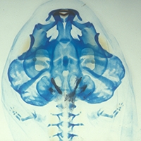
Abstract

We used RNA encoding green fluorescent protein (GFP) to study the migration and larval derivatives of cranial neural crest cells in the metamorphosing frog, Xenopus laevis. GFP provides an intrinsic cell-lineage marker that is retained after cell division. Moreover, because GFP label introduced at the one-cell stage continues to be expressed well after hatching, it offers a reliable and effective method for assessing the embryonic derivation of many larval, and possibly even adult, tissues in amphibians as well as other vertebrates. Basic patterns of cranial neural crest migration and derivation in X. laevis defined using GFP (including contributions to many larval cranial cartilages) arc similar to those documented in previous studies that used conventional vital stains, lineage markers, and ablation techniques. However, preliminary results also suggest the neural crest derivation of additional components of the larval anuran head, e.g., cranial bone, whose embryonic origins have proven much more difficult to resolve with other methods.

1999
Hanken, J., Wake, D.B. & Freeman, H.L., 1999. Three new species of minute salamanders (Thorius : Plethodontidae) from Guerrero, Mexico, including the report of a novel dental polymorphism in urodeles. Copeia , pp. 917-931.Abstract

Three new species of minute lungless salamanders of the Mexican genus Thorius (Plethodontidae) are described from montane forests in the Sierra Madre del Sur of Guerrero. Each species is distinguished from congeners by a combination of body size, external morphology, osteology, dental traits, and proteins. Thorius omiltemi and T. grandis are among the largest species within the genus; standard length (SL) approaches or exceeds 30 mm in many adults. Thorius infernalis is much smaller (SL < 19 mm). Adult T. grandis display an extreme, unique sexual dimorphism involving the presence/absence of maxillary teeth and several related features of cranial osteology. Protein (allozyme) data for T. omiltemi and T. grandis reveal substantial levels of genetic differentiation relative to species in Veracruz, Puebla, and Oaxaca. Comparable genetic data are unavailable for T. infernalis, The three species collectively define a broad elevational range, from high elevation T. omiltemi and T. grandis (2200-2700 m and 2495-3360 m, respectively) to lower montane T. infernalis (1140 m), Description of several additional species of plethodontid salamanders from central montane Guerrero underscores the region's rich herpetological diversity, which includes many endemic species of both amphibians and reptiles.

PDF

Pages