Citation:
| 272 KB |
Date Published:
MayAbstract:
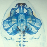
The neural crest is a population of multipotent stem cells unique to vertebrates. In the head, cranial neural crest (CNC) cells make an assortment of differentiated cell types and tissues, including neurons, melanocytes, cartilage, and bone. The earliest understanding of the developmental potentiality of CNC cells came from classic studies using amphibian embryos. Fate maps generated from these studies have been largely validated in recent years. However, a fate map for the most late-developing structures in amphibians, and especially anurans (frogs), has never been produced. One such tissue type, skull bone, has been among the most difficult tissues to study due to the long time required for its development during anuran metamorphosis, which in some species may not occur until several months, or even years, after hatching. We report a relatively simple technique for studying this elusive population of neural crest-derived osteogenic (bone-forming) cells in Xenopus laevis by using fluorescently labeled dextran conjugates. (C) 2004 Wiley-Liss, Inc.
Notes:
816FMTimes Cited:22Cited References Count:38