Publications

1995
Moury, J.D. & Hanken, J., 1995. Early cranial neural crest migration in the direct-developing frog, Eleutherodactylus coqui. Acta Anatomica , 153 , pp. 243-253.Abstract

Direct development is a common reproductive mode in Living amphibians characterized by absence of the free-living, aquatic larval stage. In Eleutherodactylus, a species-rich genus of New World frogs, evolution of direct development from the ancestral biphasic ontogeny is correlated with a comprehensive modification in embryonic cranial patterning, including the loss of many larval-specific components and the precocious formation of many adult (postmetamorphic) structures, We use scanning electron microscopy (SEM) to examine the emergence and early migration of cranial neural crest cells in Eleutherodactylus coqui to begin to assess the possible role of the neural crest in mediating these evolutionary changes. As in metamorphosing frogs, cranial crest cells emerge prior to neural fold closure and assemble into three streams: rostral, rostral otic, and caudal otic. These streams contribute to the face and first visceral (mandibular) arch, to the second (hyoid) arch, and to posterior (branchial) arches, respectively. Rostrocaudal position, morphology, and/or migration patterns distinguish subpopulations of cells within the rostral stream and caudal otic stream. With the possible exception of the small size of the rostral otic and caudal otic streams, evolution of direct development in E. coqui has not altered basic patterns of neural crest emergence or early migration as assessed by SEM. Lf observed evolutionary changes in embryonic cranial patterning are mediated by the neural crest, then they likely involve later aspects of crest migration or more subtle features related to pattern formation such as cell behavior and commitment, or gene expression.

PDF
1994
Hanken, J. & Wake, D.B., 1994. Five New Species of Minute Salamanders, Genus Thorius (Caudata, Plethodonitdaee, from Northern Oaxaca, Mexico. Copeia , pp. 573-590.Abstract

Five new species of diminutive salamanders of the endemic Mexican genus Thorius (Plethodontidae) are described from the Sierra de Juarez in northern Oaxaca. The species are diagnosed by adult body size, external proportions, dentition, osteology and coloration. The three species that have been studied using protein electrophoresis are genetically unique; all differ from T. macdougalli, the only species of the genus previously known from these mountains. Each of the six species studied has distinct geographic and elevational ranges, and there is a complex pattern of geographic overlap and replacement. As many as three species co-occur locally at elevations up to 2955 m on Cerro Pelon, and each species is sympatric with at least one other. One species descends to approximately 800 m, which is the lowest known elevational record for the genus. The new taxa include the full size range of the genus, with two large and three small species.

PDF
Seufert, D.W., Hanken, J. & Klymkowsky, M.W., 1994. Type II Collagen Distribution during Cranial Development in Xenopus Laevis. Anatomy and Embryology , 189 , pp. 81-89.Abstract

Epithelially expressed type II collagen is thought to play a prominent role in the embryonic patterning and differentiation of the vertebrate skull, primarily on the basis of data derived from amniotes. We describe the spatiotemporal distribution of type II collagen in the embryonic head of the African clawed frog, Xenopus laevis, using whole-mount and serial-section immunohistochemical analysis. We studied embryos spanning Nieuwkoop and Faber (1967) stages 21-39, a period including cranial neural crest cell migration and ending immediately before the onset of neurocranial chondrogenesis. Xenopus displays a transient expression of type II collagen beginning at least as early as stage 21; staining is most intense and widespread at stages 33/34 and 35/36 and subsequently diminishes. Collagen-positive areas include the ventrolateral surface of the brain, sensory vesicles, notochord, oropharynx, and integument. This expression pattern is similar, but not identical, to that reported for the mouse and two bird species (Japanese quail, domestic fowl); thus epithelially expressed type II collagen appears to be a phylogenetically widespread feature of vertebrate cranial development. Consistent with the proposed role of type II collagen in mediating neurocranial differentiation, most collagen-positive areas lie adjacent to subsequent sites gf chondrogenesis in the neurocranium but not the visceral skeleton. However, much of the collagen is expressed after the migration of cranial neural crest, including presumptive chondrogenic crest, seemingly too late to pattern the neurocranium by entrapment of these migrating cells.

PDF
1993
Hanken, J., 1993. Adaptation of bone growth to miniaturization of body size. In B. K. Hall, ed. Bone, Volume 7: Bone Growth - B. Boca Raton. Boca Raton: CRC Press, pp. 79-104. PDF
Hall, B.K. & Hanken, J., 1993. Bibliography of skull development: 1937-1989. In J. Hanken & B. K. Hall, ed. The Skull: Vol. 1, Development. Chicago. Chicago: University of Chicago Press, pp. 378-577.
Hanken, J. & Hall, B.K., 1993. Mechanisms of skull diversity and evolution. In J. Hanken & B. K. Hall, ed. The Skull: Vol. 3, Functional and Evolutionary Mechanisms. Chicago. Chicago: University of Chicago Press, pp. 1-36. PDF
Hanken, J. & Hall, B.K. ed., 1993. The Skull: Vol. 1, Development; Vol. 2, Patterns of Structural and Systematic Diversity; Vol. 3, Functional and Evolutionary Mechanisms, Chicago: University of Chicago Press.
Hanken, J. & Thorogood, P., 1993. Evolution and Development of the Vertebrate Skull - the Role of Pattern Formation. Trends in Ecology & Evolution , 8 , pp. 9-15.Abstract

The vertebrate skull is anatomically complex and phylogenetically diverse; it presents unique opportunities to examine the role of developmental processes in evolutionary change. Previous studies have largely examined phylogenetic trends in tissue composition or change in the timing of developmental events (heterochrony). Additional important insights may be gained if skull evolution and development are viewed from the standpoint of pattern formation. Contemporary models of pattern formation offer the possibility of linking developmental mechanisms of cranial morphogenesis from the level of genes, through cell biology, to adult form.

PDF
Hanken, J. & Wake, D.B., 1993. Miniaturization of Body Size: Organismal Consequences and Evolutionary Significance. Annual Review of Ecology and Systematics , 24 , pp. 501-519.Abstract

Miniaturization, or the evolution of extremely small adult body size, is a widespread phenomenon in animals. It has important consequences for both organismal biology and phyletic diversification above the species level. The miniaturized phenotype is a complex combination of ancestral and derived traits, including reduction and structural simplification, increased variability, and morphological novelty. Many features likely represent secondary consequences of size decrease, which may be the result of selection primarily for small body size or some related attribute such as life history characteristics. In some cases, miniaturization has resulted in novel bauplans associated with the origin of higher taxa. Evaluation of causes and consequences of miniaturization should consider obvious features of physical size as well as less obvious, but biologically important, features such as genome and cell size.

PDF
Hanken, J., 1993. Model Systems Versus Outgroups: Alternative Approaches to the Study of Head Development and Evolution. American Zoologist , 33 , pp. 448-456.Abstract

There is widespread recognition of a recent coming together of developmental and evolutionary biology in the study of problems of mutual interest. Contemporary studies into the development and evolution of the head largely comprise two parallel approaches, or research strategies: the model systems approach and the comparative approach. The two strategies share the same general goal-greater understanding of cranial development and evolution-but typically emphasize different problems, ask different questions, and employ different methods, reflecting the contrasting backgrounds and biases of each group of investigators; there has been relatively little true synthesis. Each strategy is making important and valid contributions, but both have limitations. Resolution. of many fundamental and long-standing problems in cranial development and evolution will require a combined approach that incorporates the technical and conceptual strengths of each discipline.

PDF
1992
Thorogood, P. & Hanken, J., 1992. Body building exercises. Curr. Biol. , 2 , pp. 83-85. PDF
Hanken, J., et al., 1992. Cranial Ontogeny in the Direct-Developing Frog, Eleutherodactylus coqui (Anura, Leptodactylidae), Analyzed Using Whole-Mount Immunohistochemistry. Journal of Morphology , 211 , pp. 95-118.
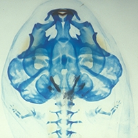
Abstract

Direct development in amphibians is an evolutionarily derived life-history mode that involves the loss of the free-living, aquatic larval stage. We examined embryos of the direct-developing anuran Eleutherodactylus coqui (Leptodactylidae) to evaluate how the biphasic pattern of cranial ontogeny of metamorphosing species has been modified in the evolution of direct development in this lineage. We employed whole-mount immunohistochemistry using a monoclonal antibody against the extracellular matrix component Type II collagen, which allows visualization of the morphology of cartilages earlier and more effectively than traditional histological procedures; these latter procedures were also used where appropriate. This represents the first time that initial chondrogenic stages of cranial development of any vertebrate have been depicted in whole-mounts.Many cranial cartilages typical of larval anurans, e.g., suprarostrals, cornua trabeculae, never form in Eleutherodactylus coqui. Consequently, many regions of the skull assume an adult, or postmetamorphic, morphology from the inception of their development. Other components, e.g., the lower jaw, jaw suspensorium, and the hyobranchial skeleton, initially assume a mid-metamorphic configuration, which is subsequently remodeled before hatching. Thirteen of the adult complement of 17 bones form in the embryo, beginning with two bones of the jaw and jaw suspensorium, the angulosplenial and squamosal. Precocious ossification of these and other jaw elements is an evolutionarily derived feature not found in metamorphosing anurans, but shared with some direct-developing caecilians. Thus, in Eleutherodactylus cranial development involves both recapitulation and repatterning of the ancestral metamorphic ontogeny. These modifications, however, are not associated with any fundamental change in adult morphology and cannot at this time be causally linked to the evolutionary success of this extraordinarily speciose genus.

PDF
Hanken, J., 1992. Life History and Morphological Evolution. Journal of Evolutionary Biology , 5 , pp. 549-557. PDF
Trueb, L. & Hanken, J., 1992. Skeletal Development in Xenopus laevis (Anura: Pipidae). Journal of Morphology , 214 , pp. 1-41.Abstract

Postembryonic skeletal development of the pipid frog Xenopus laevis is described from cleared-and-stained whole-mount specimens and sectioned material representing Nieuwkoop and Faber developmental Stages 46-65, plus postmetamorphic individuals up to 6 months old. An assessment of variation of skeletogenesis within a single population of larvae and comparison with earlier studies revealed that the timing, but not the sequence, of skeletal development in X. laevis is more variable than previously reported and poorly correlated with the development of external morphology. Examination of chondrocranial development indicates that the rostral cartilages of X. laevis are homologous with the suprarostral cartilages of non-pipoid anurans, and suggests that the peculiar chondrocranium of this taxon is derived from a more generalized pattern typical of non-pipoid frogs. Derived features of skeletal development not previously reported for X. laevis include 1) bipartite formation of the palatoquadrate; 2) precocious formation of the adult mandible; 3) origin of the angulosplenial from two centers of ossification; 4) complete erosion of the orbital cartilage during the later stages of metamorphosis; 5) development of the sphenethmoid as a membrane, rather than an endochondral bone; and 6) a pattern of timing of ossification that more closely coincides with that of the pelobatid frog Spea than that recorded for neobatrachian species.

PDF
1991
Hanken, J. & Wake, M.H. ed., 1991. Symposium: Experimental approaches to the analysis of form and function. Amer. Zool. , 31 , pp. 601-756. PDF
Wake, D.B., et al., 1991. Development and Evolution - the Emergence of a New Field. In E. C. Dudley, ed. Unity of Evolutionary Biology: The Proceedings of the Fourth International Congress of Systematic and Evolutionary Biology. Portland. Portland: Dioscorides Press, pp. 582-588. PDF
Hanken, J. & Wake, M.H., 1991. Introduction to the Symposium: Experimental Approaches to the Analysis of Form and Function. American Zoologist , 31 , pp. 603-604. PDF
Klymkowsky, M.W. & Hanken, J., 1991. Whole-Mount Staining of Xenopus and Other Vertebrates. In B. K. Kay & H. B. Peng, ed. Xenopus laevis: Practical Uses in Cell and Molecular Biology. New York. New York: Academic Press, pp. 419-441. PDF
1990
Hanken, J., 1990. Review of Thomson, K.S., Morphogenesis and Evolution. Copeia , 1990 , pp. 247-248. PDF
Dinsmore, C.E. & Hanken, J., 1990. Amputation Level-Dependent Patterning in Urodele Limb Regeneration. Journal of Morphology , 205 , pp. 77-84. PDF

Pages